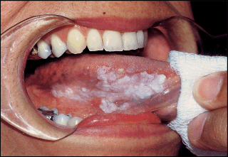
Erythroleukoplakia of the Oral Mucosa
A biopsy of a clinical erythroleukoplakia is made to determine whether
dysplasia or invasive carcinoma is present. Because a biopsy is an
invasive procedure that may cause inflammatory lymphadenopathy, which
would confuse a later TNM (primary tumor, regional nodes, and
metastases) classification if carcinoma is found, a brief clinical
staging using the TNM classification is done before the biopsy. The
biopsy may be taken using local anesthesia. However, a nerve block or
field block technique is recommended over local infiltration. Although
injecting local anesthetic into the lesion will not spread or seed tumor
cells because of the thin needle gauge, the solution volume itself will
distort tissue and create artifacts.
The most yielding areas for biopsy are the erythroplakic areas, areas of
atrophy, or areas where induration is palpated. Often multiple areas
are biopsied and, when practical, the entire clinical lesion excised (Fig 1-8).
The experienced clinician may not require biopsy site aids, but
toluidine blue may be used to determine the most yielding site for an
incisional biopsy. This aid may be of particular value in the biopsy of a
clinically homogenous lesion. Toluidine blue is a vital dye that binds
to DNA and sulfated mucopolysaccharides in all tissues. However, because
actively replicating tissues such as dysplasias and cancers contain
elevated levels of each, the blue dye will concentrate in these tissues,
thus guiding the clinician to the best site for biopsy (Figs 1-9a and 1-9b).
The technique uses a 1% aqueous solution of toluidine blue, which is
applied to the lesion and allowed to remain for 1 minute. It is then
"decolorized" with 1% acetic acid. The areas of persistent toluidine dye
staining are recommended for biopsy.
The area of biopsy should remain within the confines of the clinical
lesion. Taking a margin of normal-appearing tissue for this type of
biopsy will not assist the microscopic assessment and will only risk
extending a tumor margin into uninvolved areas. The biopsy should be
sufficiently deep to include underlying muscle. Should a carcinoma in
situ or an invasive carcinoma be found, determining the integrity of the
basement membrane and the depth of invasion, possibly into muscle
tissue, will be important.
Unless the pathologist states a preference for a different fixative, 10%
formalin (4% formaldehyde) in a neutral-buffered solution is best. If
the specimen is shipped in winter, it should be labeled, "Do not allow
to freeze," because freezing will induce artifacts.
 |
| Fig 1-8. This lesion can be excised for a complete histopathologic examination. However, if incisional biopsies are used, areas of erythroplakia, atrophy, or indurations are the best sites for sampling. |
 |
| Fig 1-9a. Suspicious floor-of-the-mouth lesion. |
 |
| Fig 1-9b. Increased uptake of 1% toluidine blue, implying a greater DNA turnover, indicates the preferred site for an incisional biopsy. |


Please keep sharing. If you want more about the best treatment for Jaw surgery specialist Kindly click the Oral and Maxillofacial Surgeon in Gurgoan
ReplyDelete